The cells shown in Image below are arthroconidia, some showing buds coming off of adjacent corners. This feature is diagnostic for Trichosporon species.
Clik here to view.

Trichosporon mucoides -Chains of arthroconidia (AC) have developed from the hyphae. Lateral blastoconidia (BC) are present, as the name implies, on the sides of the arthconidial chains or hyphae.
Clik here to view.
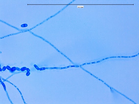
Trichosporon asahii -a hyphal element running from right to left through the center of the photo can be seen disarticulating (fragmenting) into barrel-shaped arthroconidia.
Clik here to view.

Trichosporon inkin hyphae fragmentation to arthroconidia
The yeast cells of Cryptococcus neoformans tend to be spherical, irregular in size, produce a single bud attached by a hair like connector and are surrounded by polysaccharide capsules, as illustrated in Image below.
Clik here to view.
Cryptococcus neoformans
Candida species, including C. albicans, produce single buds that tend to elongate, forming the precursor of what will be a pseudohypha. Thus, the cell shown in Image below can be suspected of being a Candida species because of the distinctly elongated bud.
Clik here to view.
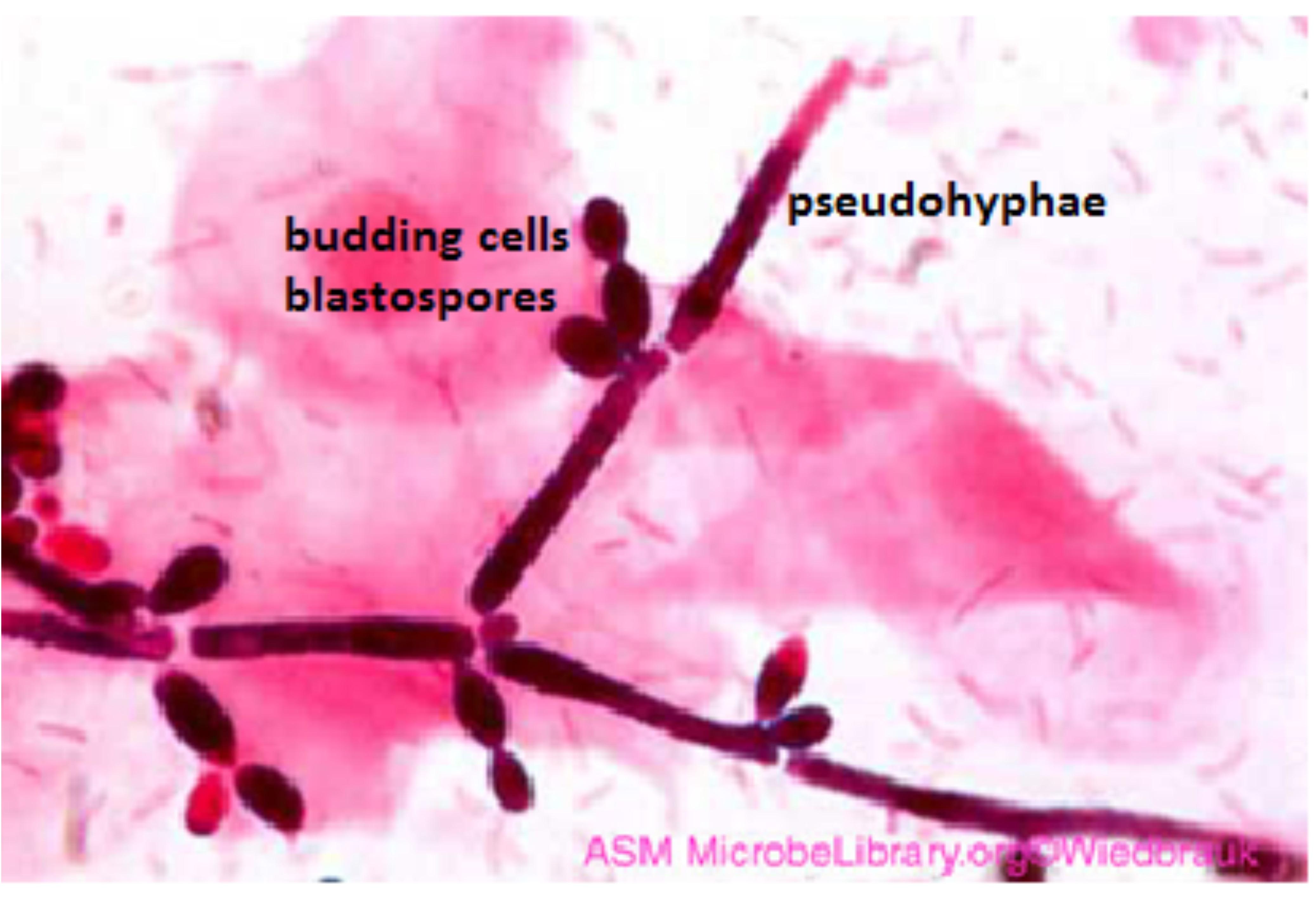
Candida sp showing budding yeasts and pseudohyphae
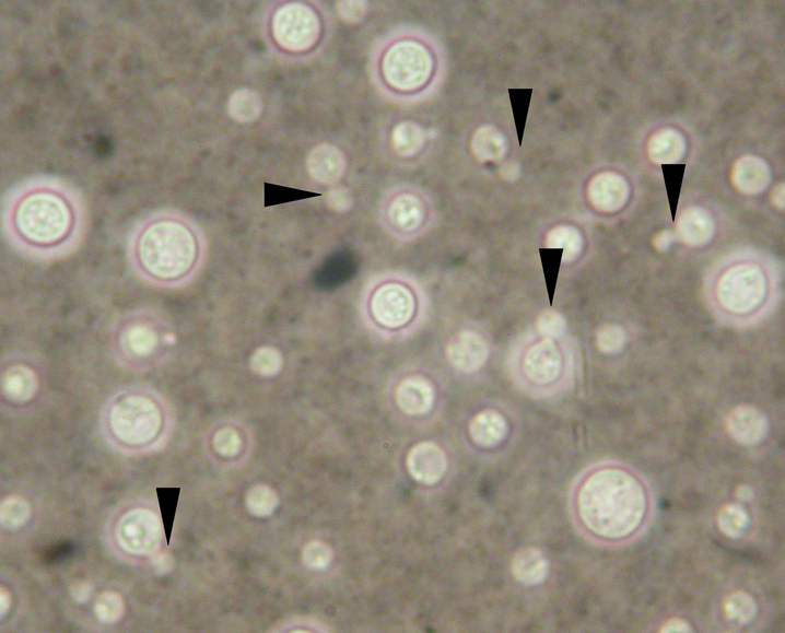
The cells shown in Image below are regular in size, have a spherical bud attached by a connector wider than seen in C. neoformans, and no surrounding capsular material. This yeast cell is most consistent with Candida (formerly Torulopsis) glabrata.
Clik here to view.

Candida glabrata yeasts
